Bacterial diseases
Bacterial diseases
Worldwide bacteria cause significant stock losses in aquaculture. Most of the bacteria that cause disease are facultative pathogens occurring naturally in the environment, only causing disease when the immune system of the host is compromised. There are some obligate bacterial pathogens (obligate = unable to live and survive without a host).
Bacteria reproduce by binary fission; where one cell divides and becomes two. Under good conditions some bacteria are able to reproduce within 20 minutes which means in only eight hours the original bacteria will have multiplied to nearly 17 million new bacteria. Some bacteria can form spores that are resistant to harsh environmental conditions.
Symptoms of bacterial fish diseases may include:
- Sudden death of many fish over a short period of time
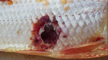
- Skin lesions, ulcers and/or fin rot
- Fish has swollen abdomen (dropsy)
- They may have exhibit pop-eye (exophthalmia)
- Cloudy eyes
- Gasping or laboured breathing
- Problems swimming or unusual swimming behaviour
Bacterial diseases - Additional link:
Most bacterial fish diseases are caused by gram negative bacteria however there are a few gram positive bacteria that cause diseases. As a general rule the gram positive bacterial fish diseases are less common but more pathogenic than most gram negative infections.
Aeromonas hydrophila (Bacterial Hemorrhagic Septicemia)
- Gram negative motile rods.
- Effects many freshwater species and usually is associated with stress and overcrowding. The clinical signs and lesions are variable. The most common finding is hemorrhage in skin, fins, oral cavity and muscles with superficial ulceration of the epidermis. Occasionally cavitary ulcers are observed. Exophthalmus and ascites are commonly observed. Splenomegaly and swollen kidneys are common. Histologically, multifocal areas of necrosis in the spleen, liver, kidney and heart with numerous rod shaped bacteria are observed.
- Diagnosis is rendered by culturing the organism from affected animals: Remember this is a common water saprophyte with a great variation in virulence in serotypes.
- Disease is transmitted via contaminated water or diseased fish.
Pseudomonas fluorescens
Vibrio
- Gram negative rod, lives primarily in a marine environment
- Vibrio septicemia: V. alginolyticus / V. anquillarum / V. salmonicida 1. Septicemia has similar lesions to Aeromonas hydrophila.
- Ulcer Disease of Damselfish: V. damsela 1. Deep skin ulcers and necrotizing myositis.
- Vibrio salmonicida: Hitra disease or Cold water vibriosis
2. See hemorrhage in the skin of the tail and fins, ulceration of the skin, hemorrhage in the muscles and serosal surfaces. The spleen may be enlarged and bright red. Histologically may see necrosis of the liver, kidney, spleen and occasionally the gut mucosa.
2. Lesions similar to Aeromonas salmonicida.
Edwardsiella tarda (Edwardsiella septicemia)
- Gram negative motile pleomorphic curved rod
- The disease affects primarily channel catfish but also observed in goldfish, golden shiners, largemouth bass, and the brown bullhead. This organism is the most serious disease involving the eel culture of Asia.
- The lesions are similar to A. hydrophila with small cutaneous ulcers and hemorrhage observed both in the skin and muscle. Muscle lesions often develop into large gas filled (malodorous) cavities. Diseased fish lose control over the posterior half of their body yet continue to feed.
- Gram negative motile pleomorphic curved rod
- Disease affects primarily fingerlings and yearling catfish
- Clinical signs of enteric septicemia of catfish closely resemble those of other systemic bacterial infections.
The most characteristic external lesion is the presence of a raised or open ulcer on the frontal bone of the skull between the eyes (Hole in the head disease). - Gram negative non-motile short rod
- Bacteria affect primarily salmonids but other freshwater fish can be affected.
- Clinically the disease may present as a septicemia with hemorrhage in the muscles and other sites. The major lesion is a subcutaneous swelling that often causes an ulcerative dermatitis. In chronic disease these lesions may cavitate into the adjacent musculature. In the septicemic disease, there is splenomegaly, ascites, and swelling of the kidneys. Histologically, there is necrosis of the affected tissue with abundant colonies of bacteria and few inflammatory cells due to the bacteria's leukocytolytic exotoxin.
- The disease is transmitted by contact with diseased fish, contaminated water, fomites, and infected eggs.
- Gram negative motile rod.
- The bacteria affect salmonids; rainbow trout are the most susceptible.
- Clinically this disease manifests itself as a septicemia with exophthalmus, ascites, and hemorrhage and ulceration of the jaw, palate, gills and operculum. Hemorrhage of the musculature and serosal surfaces of the intestines, splenomegaly, and kidney swelling are common. Histologically numerous bacterial colonies admixed with inflammatory cells are observed in many areas of necrosis involving the liver, spleen and kidney.
- The disease is transmitted by contact with diseased or carrier fish, and contaminated water. Bacteria persist in asymptomatic non-salmonid fish and in some birds.
- Beta-hemolytic Streptococcus (Note: Beta hemolysin may not be present in culture media in all cases leading to the possible believes that this bacteria is a non-pathogen).
- Disease of tilapia, hybrid striped bass and rainbow trout.
- Major problem in the tilapia industry. Streptococcus iniae presents either as an acute fulminating septicemia or in a chronic form limited primarily to the central nervous system. The septicemic form may present with hemorrhage of the fins, skin, and serosal surfaces. Ulcers may appear. Microscopically, one observes a meningoencephalitis, polyserositis, epicarditis, myocarditis and/or cellulitis. Cocci/diplococci are present in the inflammation. In the chronic form, granulomas or granulomatous inflammation are evident in the liver, kidney, and brain (meningoencephalitis). In the chronic disease, the brain is the best organ to culture.
- Streptococcus iniae is a problem primarily of closed recirculating culture system. Probably associated with overcrowding and poor water quality - high nitrates. Depopulation, disinfection and restocking with disease free fish are the best means of elimination of the organism.
- The bacteria are known to be a zoonotic agent. Individuals who have handled infected fish have developed cellulitis of the hands and endocarditis.
- Gram negative slender rods (3-8 microns)
- The disease is a serious disease of young salmonids, catfish and many other fish.
- This is a highly communicable disease. Lesions usually first appear as small white spots on the caudal fin and progresses towards the head. The caudal fin and anal fins may become severely eroded. As the disease progresses, the skin is often involved with numerous gray white ulcers. Gills are a common site of damage and may be the only affected area. The gill lesions are characterized by necrosis of the distal end of the gill filament that progresses basally to involve the entire filament.
- Flexibacter columnaris infections are frequently associated with stress conditions. Predisposing factors for Columnaris disease are high water temperature (25°C-32°C.), crowding, injury, and poor water quality (low oxygen and increased concentrations of free ammonia).
- Flexibacter maritimus: cause similar problems in salt-water environment.
- Flexibacter psychrophilus causes Cold Water Disease or Peduncle disease. Fish develop dark skin, hemorrhage at the base of fins, and anemia with pale gills with increase mucus. Hemorrhage into the muscles is common. Periostitis of cranial and vertebral bones is common in chronic cases. A chronic meningoencephalitis occasionally is observed with abnormal and erratic swimming.
- Bacterial gill disease is caused by a variety of bacteria. Flexibacter columnaris, Flexibacter psychrophilus, Cytophagy psychrophila and various species of Flavobacterium (all are gram negative rods) are the primary bacteria involved in this disease.
- Fry are the most susceptible to the disease, however, all ages may be affected. Clinically the fish become anorectic, and face the water current. Prominent hyperplasia (mucus and epithelial) of the gills is evident on gross and microscopic examination. Microscopically one observes proliferation of the epithelium that result in clubbing and fusion of the lamella. Necrosis of the gill lamella occurs in serious cases.
- Overcrowding, accumulation of metabolite waste products (particularly ammonia), organic matter in the water, and an increase in water temperature may all be predisposing factors.
- Gram negative filamentous bacteria
- Occurs primarily in rainbow trout fry. Fish develop abdominal distention, exophthalmus, increased pigmentation, lethargy, loss of balance, pale gills and occasional cutaneous ulcers and necrosis of tail fins. Epidermal hyperemia and increase mucus secretions are common. Splenomegaly and hepatomegaly are common with multifocal necrosis of the liver spleen and kidney.
- Transmission is believed to be by direct contact with contaminated water and is an indication of poor water quality and overcrowding.
- Gram positive nonmotile diplobacillus.
- This is a serious disease of salmonids. Brook trout are the most severely affected species.
- The disease follows a slow course with clinical signs not present until the fish is well grown. The fish may exhibit exophthalmus, skin darkening, and hemorrhage at the base of the fins. Cutaneous vesicles and ulcers may develop in mature trout "spawning rash". Abscesses, cavitation and contraction of muscles is occasionally observed. Splenomegaly and swelling of the kidney and liver with abundant ascites fluid is commonly observed. The large swollen kidney and spleen have numerous white nodules visible in the parenchyma. Numerous granulomas (containing gram positive bacteria) are observed in the kidney and may be also present in the spleen, heart and liver.
- Transmission of the disease is believed to be via direct contact with contaminated fish. It is believed that the organism enters through the epidermis and then becomes a systemic disease.
- Gram positive, acid fast rods (M. marinum, M. chelonei and M. fortuitum are the most common Mycobacterium species involved.)
- All species of fish are affected. This disease affects both saltwater and freshwater fish raised for food as well as aquariums (up to 10 to 25% of pen raised fish).
- Clinical signs of tuberculosis are quite variable. The most common signs are anorexia, emaciation, vertebral deformities, exophthalmus, and loss of normal coloration. Numerous variably sized granulomas are often observed in various organs throughout the body. Often numerous acid fast bacteria are observed in the granulomas.
- The aquatic environment is believed to be the source of initial infection with fish becoming infected by ingestion of bacterial contaminated feed or debris. Once an aquarium is infected with this disease, it is difficult to remove except by depopulation of the aquarium and disinfecting the tank. Remember this is a zoonotic disease (atypical mycobacteriosis).
- Atypical mycobacteriosis may manifest itself as a single cutaneous nodule on the hand or finger or may produce a regional granulomatous lymphadenitis of the lymphatics near the original nodule. Occasional local osteomyelitis and arthritis may also occur.
- Gram positive filamentous rod (weakly acid fast positive)
- The organism is a problem with mostly aquarium fish. However, it is occasionally observed in cultured salmonids.
- Clinically this is a chronic disease characterized by raised granulomatous masses in the mouth, jaw, gills and skin (The mouth and jaw are the most common sites). Dermal masses eventually ulcerate. Numerous white raised nodules (granulomas) are often observed in the viscera.
- The exact route of transmission is unknown. However, it is felt that entry through wounds and abrasions is the most common source of infection. (Ingestion of the bacteria has been known to cause the disease.)
- Gram negative rods
- Usually a problem for individual fish. This disease is a cause of concern to primarily hobbyist and producers of ornamental fish. (Mollie granuloma, Mollie madness, Mollie popeye)
- Infected fish are usually emaciated and pale. Multifocal white nodules are observed in the visceral organs, the retina and choroid and the brain. These nodules may be cystic or mineralized. Histologically the nodules are granulomas with a caseous center, a thin peripheral rim of macrophages and lymphocytes and a fibrous capsule.(Must be differentiated from Mycobacterium)
- The mode of transmission is unknown.
- Obligated intercellular parasite. Organisms stain red with Macchiavello stain.
- These organisms have been observed in many species of fresh water and marine fish. Mortality occurs most commonly in heavily infected juvenile fish.
- Clinically infected fish may be asymptomatic or show respiratory distress or excessive mucus secretions. Multiple white cysts are observed on the gill lamella and skin. Histologically, the cyst consists of distended epithelial cells with numerous basophilic organisms.
- The means of transmission is unknown.
Edwardsiella ictaluri (Enteric septicemia of catfish)
Aeromonas sp. (Furunculosis, Ulcerative disease)
Yersinia ruckeri (Enteric red mouth)
Streptococcus iniae
Flexibacter columnaris (Columnaris disease or Saddleback disease)
Bacterial Gill Disease:
Cytophaga psychrophila (Rainbow Trout Fry Anemia)
Renibacterium salmoninarum (Bacterial Kidney Disease)
Mycobacterium species (Tuberculosis)
Nocardia sp.
Flavobacterium sp.
Epitheliocystis (Chlamydial infection)
- Address
- Neospark Drugs and Chemicals Private Limited Corporate Center,
241, B.L. Bagh, Panjagutta,
Hyderabad- 500 082,
Telangana, India.
- Product Groups
- Poultry
- Large Animal
- Aquaculture
- Feed
© 2026 Neospark Drugs and Chemicals Private Limited